In 2019, Jia Liu, PhD, didn’t know where his own pancreas was. He was just starting his assistant professorship at Harvard, building stretchable mesh electronics for brain and heart organoids. Pancreatic cells, let alone the pancreas, were not on his radar. That is, until Juan Alvarez-Dominguez, PhD, then a postdoctoral student in the lab of Douglas Melton, PhD, heard Liu give a talk.
Melton pioneered the differentiation of insulin-producing beta cells from human stem cells, giving Alvarez-Dominguez unparalleled training under one of the field’s founders. He also knew something most engineers didn’t: Pancreatic beta cells and brain neurons share an evolutionary ancestor, express the same ion channels, and release their cargo through the same electrical machinery. If electronics could read neurons, they could read beta cells.
After Liu’s talk, Alvarez-Dominguez met with Melton. And Melton, whose decades of work on beta cell differentiation now underpins Vertex Pharmaceuticals’ islet cell therapy program, then asked to meet with Liu.
“You cannot say no when [Douglas Melton] wants to meet with you,” Liu said.
During the meeting, Melton described a problem to Liu: During the early Vertex transplant trials, when grafts failed in some patients, the clinical team had no reliable way to know if the islet cells were even alive. Cells may look healthy under a microscope, but a visual check does not give the full picture of functionality. No one was measuring whether the cells were firing, responding to glucose, or producing insulin in real time.
A collaboration was born.
Now, 6 years later, that collaboration has produced a fascinating study, co-led by Liu and Alvarez-Dominguez. They were able to coax stem cell-derived islet cells to coalesce around a microscopic, flexible electronic sensor and function normally while this “cyborg” organoid transmits troves of new data on how islet cells live and mature.
120 Days vs 12 Days
The maturation problem sits at the heart of stem cell-derived islet therapy. Islet cells from deceased donors, when transplanted, can start working in about 12 days. But stem cell-derived islets take 10 times longer, closer to 120 days, Alvarez-Dominguez said. For patients injecting insulin every day, every extra day waiting is another day of finger sticks, insulin doses, and the risk of a hypoglycemic episode that can land someone in the emergency room.
Nobody fully understands why lab-grown islet cells remain functionally immature in cell culture but mature after transplantation. “If we did, you’d have a lot of papers out there saying, ‘here is a protocol that makes maximally mature beta cells,’” said Jeffrey R. Millman, PhD, an associate professor of medicine and biomedical engineering at Washington University in St Louis, who was not involved in the study.
Researchers have spent years trying to close this gap, but the gains have been modest. “Maybe 1% closer to full maturation,” Millman said, “but you’re still ignoring 99% of the path.”
The Signal Problem
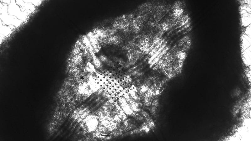
Liu and Alvarez-Dominguez’s approach was to embed ultrathin, stretchable mesh electronics into stem cell-derived pancreatic organoids during the earliest stages of development. The mesh — barely visible to the naked eye, with wavy interconnects designed to stretch — is laid flat and seeded with stem cells. Within 48 hours, the cells begin clustering into a three-dimensional organoid, and as they fold inward, they drag the electronics with them.
The result is a living tissue with sensors woven throughout its interior, like rebar inside concrete.
Yet for months, the electronics captured almost nothing useful. Pancreatic alpha and beta cells are only about 10 micrometers across — five times smaller than heart cells, with none of the long projections that make neurons easy to record. All the team could pick up was crowd noise.
“We initially thought we couldn’t isolate any sharper signal,” Liu said.
A student figured out the solution. By integrating the electronics at an earlier differentiation stage, before the cells had fully committed to their identities, the cells could form tight junctions around the sensors as they matured together, transforming the quality of the signal into something much more useful.
When the team compared organoids with electronics to organoids without, the cells were indistinguishable, as if the electronics were invisible to the biological reality around it.
“This device is highly noninvasive. It grows together with the cell very well. It’s a passive measurement, so it’s not interrupting the tissue,” Liu said.
Two States
The team then did something that had never been achieved in pancreatic tissue: They simultaneously distinguished alpha and beta cell electrical activity at single-cell resolution within intact three-dimensional organoids and tracked it for months.
No previous method could do what a clinician needs: watch alpha and beta cells simultaneously, within intact tissue, over the weeks to months that maturation happens.
The recordings revealed two distinct populations within alpha and beta cells. One group stayed quiet until glucose arrived, then fired strongly — behaving like mature, adult islet cells that wait for the right signal. The other group fired constantly, even when glucose was low, behaving like fetal cells that haven’t yet learned when to stay silent.
You might expect the immature cells to gradually learn discipline — to stop firing out of turn. Instead, every cell in the organoid simply turned up its volume. The well-behaved ones fired more forcefully when glucose arrived. The ones beating to the tune of their own drum, so to speak, also ramped up.
Until now, scientists assumed that what changed during maturation was the cell’s response to its own electrical signal, specifically, how sensitive it became to calcium, the molecule that ultimately triggers insulin release. The electrical firing was thought to stay the same. But this team found the opposite: What changes is the firing itself. Mature cells set a higher bar for when they fire in the first place. The gatekeeper, not the gate, is what matures.
Bozhi Tian, PhD, a professor of chemistry at the University of Chicago who develops bioelectronic tissue interfaces and was not involved in the study, called the work “truly groundbreaking,” noting that single-cell electrophysiology within a 3D organoid adds “temporal precision and cell-type specificity at the level of excitable dynamics” beyond what population-averaged assays can provide.
In practical terms, that means clinicians could one day evaluate whether a batch of lab-grown islets is ready for transplantation the way a cardiologist reads an EKG — by listening to the electrical signature of individual cells, not just measuring what the tissue secretes into a dish.
A Sleep Schedule for an Organoid
After 2 months of culture, the team gave the organoids something they’d never had: a daily schedule. For 4 days, they cycled between periods of glucose stimulation and rest.
“Functional maturation is not a time-dependent process,” Alvarez-Dominguez said. “And if it’s not a function of time, you can modulate the context to accelerate it.”
The entrained organoids soon developed daily rhythms in the shape of each electrical pulse, oscillating with time of day. Control organoids without the schedule lost electrical activity entirely within two days. After 4 days of training, the rhythm persisted on its own.
The team then tried something more direct: delivering fixed electrical pulses through the embedded electronics. Cells that should respond to high glucose — beta cells — became more responsive to high glucose. Cells that should respond to low glucose — alpha cells — became more responsive to low glucose. Neither group changed its baseline behavior. The stimulation sharpened what each cell was already supposed to do, without forcing anything inappropriate.
Millman urged caution. “There are just not a lot of data on that,” he said of the stimulation results. “It’s still quite speculative.” His bar: show these cells outperform a modern protocol in a diabetic mouse model. “Maybe a lower dose, maybe a faster cure.”
Two Paths Forward
The team’s vision extends in two directions. The nearer goal is what Tian described as a “training scaffold,” in which electronics condition cells toward maturity in the lab, then dissolve before transplantation. The further goal is to send the electronics into the body alongside the cells, as a subcutaneous device resembling a continuous glucose monitor, wirelessly synced to a phone.
If the cells stop working, whether from immune attack or metabolic stress, the device could detect the silence and stimulate them back online.
“If the cells are alive, they produce electricity,” Alvarez-Dominguez said. “If they’re not, they don’t.”
The platform isn’t ready for patients, obviously. It monitors only a few hundred cells out of roughly a million, and recordings last five minutes, insufficient for the continuous monitoring clinicians need.
Liu called the limitation a “purely engineering problem” that can be solved: His team is building wireless modules to tackle it, though progress slowed when federal funding was interrupted for several months.
Asked what this could mean for someone with type 1 diabetes today, Liu was honest: Regulatory approval for implantation is years away. But the technology could be used now to screen lab-grown islets before transplantation, sorting the mature from the immature, so patients receive a better product.
Alvarez-Dominguez was more direct. “There is no technical impediment for us to try these applications,” he said. “It’s just a matter of integrating the parts and getting funded.”
Tian and Millman reported no relevant financial relationships. Disclosure information for study authors is available in the original study publication.
